1. Human eyes are a pair of organs located in sockets of the skull called orbits.
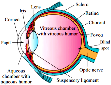
2. Eyeball is spherical and has three layers.
3. Sclera is the outer layer of dense connective tissue with anterior transparent cornea.
4. Choroid is the middle layer. It is bluish in colour containing many blood vessels. The anterior region is thick and forms the ciliary body. Posterior 2/3rd region is thinner.
5. Iris is the forward segment of the ciliary body which is pigmented and opaque. This part is the visible coloured portion of the eye.
6. Lens is present anteriorly inside the iris and is held in position by the ligaments of ciliary body.
7. The aperture surrounded by the iris in front of the lens is known as pupil. The movement of the pupil is regulated by the muscle fibres of iris.
8. The innermost layer of the eye is the retina having three sub-layers formed by ganglion cells, bipolar cells and photoreceptor cells, which are sensitive to light.
9. There are two types of photoreceptor cells, viz. rods and cones containing light sensitive proteins. They are termed as photo pigments, rhodopsin which is a derivative of vitamin A (in rods) and iodopsin (in cones).
10. The cones are responsible for daylight or photopic vision and colour vision. The rods function in dim light giving scotopic vision.
11. The cones are of three types, each containing its own characteristic photopigments that respond to red, green and blue lights.
12. The optic nerve leaves the eye at a point slightly away from the median posterior pole of the eyeball. In this region, the rods and cones are absent therefore this region is known as blind spot. Macula lutea, a yellowish pigmented spot is present lateral to the blind spot.
13. Fovea is a central pit present beside it. Fovea is a thinned out portion of the retina where only the cones are densely packed and therefore have greatest visual acuity (resolution).
14. A space between the cornea and the lens is called aqueous chamber. It contains a thin watery fluid known as aqueous humor.