1. Take permanent slides which shows different stages of mitotic cell division from your lab kit.
2. Observe carefully under microscope.
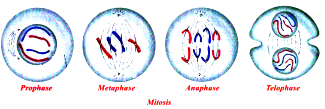
3. Draw diagrams what you observe, and compare your observations with the following chart.
| Stage |
Description |
| 1. Prophase |
1. Chromosomes contract, spiral and become visible even in light microscope and nucleoli become smaller (material to chromosomes).
2. Chromosomes split lengthwise to form chromatids, connected by centromeres.
3. Nuclear membrane disappears.
4. Centrosome, containing rod-like centrioles, divides and forms ends of spindle (probably animal cells only). (Note : No pairing of chromosomes as in meiosis). |
| 2. Metaphase |
1. Chromosomes move to spindle equator, spindle fibres attached to centromeres. |
| 3. Anaphase |
1. Centromeres split, separating the chromatids.
2. Spindle fibres attached to centromeres contract, pulling chromatids towards poles. |
| 4. Telophase |
1. Chromatids elongate, become invisible, (replication at this stage to become chromosomes).
2. Nuclear membranes form round daughter nuclei.
3. Cell membrane pinches in to form daughter cells (animals) or new cell wall material becomes laid down across spindle equator (plants).
4. Nucleus divides into two and division of cytoplasm starts. |